44 brain mri with labels
What Does a Brain MRI Show? - San Diego Health What does a brain MRI show? The answer is, unfortunately, not very. MRI scans (magnetic resonance imaging) have been around for decades, and the technology has been steadily improving. Today, a brain MRI test can identify whether or not a person has a stroke, or if the person has suffered a traumatic brain injury, or if the person is suffering ... 101 Labeled Brain Images and a Consistent Human Cortical Labeling ... Labeled anatomical subdivisions of the brain enable one to quantify and report brain imaging data within brain regions, which is routinely done for functional, diffusion, and structural magnetic resonance images (f/d/MRI) and positron emission tomography data.
Atlas of BRAIN MRI - W-Radiology Brain magnetic resonance imaging (MRI) is a common medical imaging method that allows clinicians to examine the brain's anatomy (1). It uses a magnetic field and radio waves to produce detailed images of the brain and the brainstem to detect various conditions (2).

Brain mri with labels
Cross-sectional anatomy of the brain - e-Anatomy - IMAIOS Apr 15, 2022 · Axial MRI Atlas of the Brain. Free online atlas with a comprehensive series of T1, contrast-enhanced T1, T2, T2*, FLAIR, Diffusion -weighted axial images from a normal humain brain. Scroll through the images with detailed labeling using our interactive interface. Perfect for clinicians, radiologists and residents reading brain MRI studies. 101 labeled brain images and a consistent human cortical labeling ... given how difficult it is to label brains, the mindboggle-101 dataset is intended to serve as brain atlases for use in labeling other brains, as a normative dataset to establish morphometric variation in a healthy population for comparison against clinical populations, and contribute to the development, training, testing, and evaluation of … Anatomy of the foot and ankle - MRI - e-Anatomy - IMAIOS Sep 13, 2021 · Cross-sectional anatomy: MRI of the ankle and feet. A magnetic resonance imaging (MRI) was performed on a normal subject; with spin-echo T1 weighted images and spin-echo fat-saturated proton density weighted images (3 usual planes used for osteo-articular imaging: axial, coronal, and sagittal).
Brain mri with labels. Researchers automate brain MRI image labeling, more than 100,000 exams ... Researchers have automated brain MRI image labeling, needed to teach machine learning image recognition models, by deriving important labels from radiology reports and accurately assigning them to... GitHub - yunshiuan/label4MRI: Label the brain MNI coordinate by AAL/BA ... MRI-labeling: label human brain MRI image by AAL/BA system Under the R program environment,input an MNI coordinate, output the corresponding AAL(Automated Anatomical Labeling) and BA (Brodmann area) brain region name. More importantly, if the coordinate does not match a brain region defined by AAL/BA (e.g., white matter), the package help find the closest brain region with the corresponding ... Brain MRI: What It Is, Purpose, Procedure & Results A brain MRI (magnetic resonance imaging) scan, also called a head MRI, is a painless procedure that produces very clear images of the structures inside of your head — mainly, your brain. MRI uses a large magnet, radio waves and a computer to produce these detailed images. It doesn't use radiation. Labeled MRI Brain Scans - Neuromorphometrics We can also label scans that you provide and we are very interested in labeling white matter anatomy as seen in diffusion-weighted MRI scans. If you want an aggregate version of our data, we can provide it as a probabilistic atlas. The cost to label a single scan is $2449 (USD).
Enhancing the REMBRANDT MRI collection with expert segmentation labels ... Data download. We first downloaded the pre-operative raw MRI scans from the TCIA imaging archive 10,20 for all the 130 patients including multiple series for each patient in DICOM file format 21 ... Automatic anatomical brain MRI segmentation combining label propagation ... Using the brain label data, we determined the variation of a and b depending on the registration method employed (unregistered, ... Manual and automated measurement of the whole thalamus and mediodorsal nucleus using magnetic resonance imaging. NeuroImage, 17 (2) (2002), pp. 631-642. Article Download PDF View Record in Scopus Google Scholar. › video › indexMail Online Videos: Top News & Viral Videos, Clips & Footage ... Jul 06, 2022 · Check out the latest breaking news videos and viral videos covering showbiz, sport, fashion, technology, and more from the Daily Mail and Mail on Sunday. Split-Attention U-Net: A Fully Convolutional Network for Robust Multi ... Multi-label brain segmentation from brain magnetic resonance imaging (MRI) provides valuable structural information for most neurological analyses. Due to the complexity of the brain segmentation algorithm, it could delay the delivery of neuroimaging findings. Therefore, we introduce Split-Attention …
Mail Online Videos: Top News & Viral Videos, Clips & Footage Jul 06, 2022 · Check out the latest breaking news videos and viral videos covering showbiz, sport, fashion, technology, and more from the Daily Mail and Mail on Sunday. › panoramic › indexPharmaCircle This website uses cookies to help provide you with the best possible online experience. Please read our Terms & Conditions and Privacy Policy for information about ... Videojug - YouTube Welcome to Videojug! Here you'll find the best how-to videos around, from delicious, easy-to-follow recipes to beauty and fashion tips. MRI Brain Atlas This web app Atlas is intended for veterinary students and radiologists seeking quick access to canine brain anatomy through a mobile device. Via a toggle button, either MRI images or approximately comparable Brain Transection images may be viewed with or without labels. Navigation & Labels.
Neuroradiology Cases: Brain Abscess MRI
Brain: Atlas of human anatomy with MRI - e-Anatomy - IMAIOS Sep 13, 2021 · MRI Atlas of the Brain. This page presents a comprehensive series of labeled axial, sagittal and coronal images from a normal human brain magnetic resonance imaging exam. This MRI brain cross-sectional anatomy tool serves as a reference atlas to guide radiologists and researchers in the accurate identification of the brain structures.
Ct and mri interpretation
Automated MRI image labelling processes 100,000 brain exams in under 30 ... Researchers from the School of Biomedical Engineering & Imaging Sciences at King's College London have automated brain MRI image labeling, needed to teach machine learning image recognition models,...

Bl4ck D4ys: March 2011
Brain MRI: How to read MRI brain scan | Kenhub MRI is the most sensitive imaging method when it comes to examining the structure of the brain and spinal cord. It works by exciting the tissue hydrogen protons, which in turn emit electromagnetic signals back to the MRI machine. The MRI machine detects their intensity and translates it into a gray-scale MRI image.
Images from Radiological Testing -- refried.org weblog
UCLA Brain Mapping Center - ICBM Template To view both the structural MRI and the labels launch the program typing Display icbm_template.mnc -label icbm_labels_corrected.mnc. The opacity of the labels can be set in the Colour Coding menu. The number of each label appears at the bottom left of the orthogonal views window.

ON - RADIOLOGY: Lipoma arborescens of Ankle MRI (what`s that !!!)
Brain lobes - annotated MRI | Radiology Case | Radiopaedia.org Debowski, M. Brain lobes - annotated MRI. Case study, Radiopaedia.org. (accessed on 06 Jul 2022)
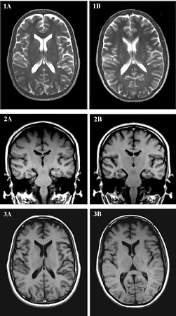
Brain MRI Comparison Images
PharmaCircle This website uses cookies to help provide you with the best possible online experience. Please read our Terms & Conditions and Privacy Policy for information about ...
MRI Procedure of Brain
MRI Brain Animated Quiz - University of Minnesota Note: spacebar toggles labels; also arrow keys do Previous/Next Sequentially click/tap: first the dot associated with a term; then, its corresponding target dot on the MRI image. If a line connection appears, your choice was correct!

RADIOLOGY: HIV Encephalopathy
Labels · ryx2/MRI-Brain-Registration · GitHub My master's project. Contribute to ryx2/MRI-Brain-Registration development by creating an account on GitHub.
Post a Comment for "44 brain mri with labels"