40 images of compound microscope with labels
Microscope Components - Science Quiz - GeoGuessr Microscope Components - Science Quiz: The most common type of modern microscope is called a compound microscope. They have two systems of lenses, one is the eyepiece and the other is comprised of one or more objective lenses. This type of microscope has become so advanced that some are capable of magnifying up to 1000 times! Microscopes are used in … Looking at the Structure of Cells in the Microscope A typical animal cell is 10–20 μm in diameter, which is about one-fifth the size of the smallest particle visible to the naked eye. It was not until good light microscopes became available in the early part of the nineteenth century that all plant and animal tissues were discovered to be aggregates of individual cells. This discovery, proposed as the cell doctrine by Schleiden and …
Food Calorimetry: How to Measure Calories in Food - Carolina.com We have the compound microscope you are looking for! Digital Microscopes. Digital microscopes are great for large classroom computer combined instruction. Students can take images, videos, and more. Stereomicroscopes. Stereomicroscopes show 3D images vs. flat images and are easier to focus and use. They are great for first tme student use.

Images of compound microscope with labels
Electron microscope - Wikipedia An electron microscope is a microscope that uses a beam of accelerated electrons as a source of illumination. As the wavelength of an electron can be up to 100,000 times shorter than that of visible light photons, electron microscopes have a higher resolving power than light microscopes and can reveal the structure of smaller objects.. Electron microscopes use shaped magnetic … (PDF) Introduction to Microscopy - ResearchGate • In compound microscope it will be i.e 10 X, f= 16 mm; 40 X, f= 4 mm; 100 X, f= 1.8 mm. • Image produced by objective lens falls on the eyepiece lens serve as objec t. • … Amazon.com: My First Lab Duo Scope Microscope - Young … Duo-Scope functions as a compound microscope and stereomicroscope in one unit. ... and experiment guide, includes 5 blank slides, 1 concavity (well) slide, 4 prepared slides, 2 bottles of stain, 5 slide labels, 5 cover glasses, 50 sheets of lens paper, 1 plastic transfer pipette, 1 plain wooden applicator, 1 cotton-tipped applicator, 1 plastic ...
Images of compound microscope with labels. Parts of Stereo Microscope (Dissecting microscope) - Rs' Science The key is presenting two images with a slightly different angle to our brain. Photo source: dailymotion. ... Compared to a compound microscope where the objectives attached to the nosepiece can be seen and identified individually (based on color bands and their respective labels), the objectives of a dissecting microscope are located in a ... Cell Types Gizmo Worksheet - StuDocu Select the MICROSCOPE tab. Introduction: Complex organisms are made up of smaller units, called cells. Most cells are too small to be seen by the naked eye. Microscopes are used to magnify small objects, so here you will use a compound light microscope to observe the cells of different organisms. What is Electron Microscopy? - UMASS Medical School Electron microscopy (EM) is a technique for obtaining high resolution images of biological and non-biological specimens. ... The TEM is analogous in many ways to the conventional (compound) light microscope. TEM is used, among other things, to image the interior of cells (in thin sections), the structure of protein molecules (contrasted by ... Multiphoton Microscopy | Nikon’s MicroscopyU The images presented in Figure 7 (a shark choroid plexus stained with fluorescein) provide a comparison of confocal and two-photon microscopy imaging quality. These images were collected at 80-micrometers below the specimen surface, which is the maximal depth allowing sufficient image contrast from this specimen utilizing confocal microscopy.
Amazon.com: My First Lab Duo Scope Microscope - Young … Duo-Scope functions as a compound microscope and stereomicroscope in one unit. ... and experiment guide, includes 5 blank slides, 1 concavity (well) slide, 4 prepared slides, 2 bottles of stain, 5 slide labels, 5 cover glasses, 50 sheets of lens paper, 1 plastic transfer pipette, 1 plain wooden applicator, 1 cotton-tipped applicator, 1 plastic ... (PDF) Introduction to Microscopy - ResearchGate • In compound microscope it will be i.e 10 X, f= 16 mm; 40 X, f= 4 mm; 100 X, f= 1.8 mm. • Image produced by objective lens falls on the eyepiece lens serve as objec t. • … Electron microscope - Wikipedia An electron microscope is a microscope that uses a beam of accelerated electrons as a source of illumination. As the wavelength of an electron can be up to 100,000 times shorter than that of visible light photons, electron microscopes have a higher resolving power than light microscopes and can reveal the structure of smaller objects.. Electron microscopes use shaped magnetic …
shikha mahajan: Compound Microscope
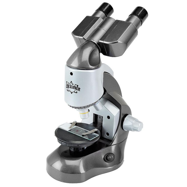
Edu Science - M1280X Ultra Student Microscope | Toys R Us Canada
Print Microbiology Lab 2 (Microscopy, Gram stain, intro to enterotube II) flashcards | Easy ...
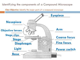
PPT - Identifying the components of a Compound Microscope PowerPoint Presentation - ID:2045335

Types of Microscope🔬| Simple-Compound-Special Microscope | - YouTube
How to Use a Compound Microscope Basic Microscopy

Microscope World Blog: Biological Microscope Magnifications

labels of a compound microscope microscope boxed - Top Label Maker

Microscope World Blog: Human Anatomy under the Microscope

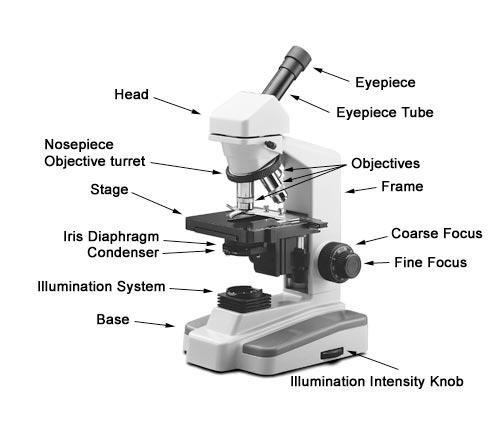
31 Compound Microscope With Label - Labels For Your Ideas
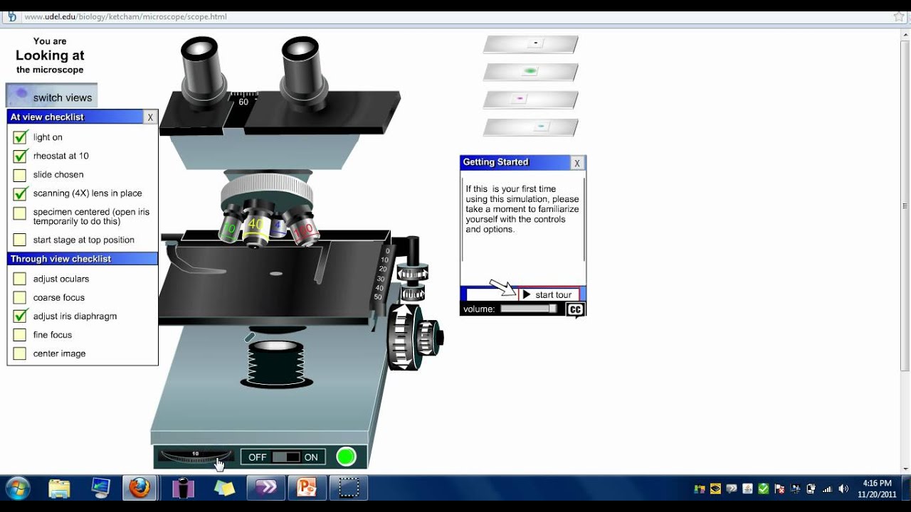
virtual microscope lab - YouTube

Microscope World Blog: Biological Microscope Magnifications
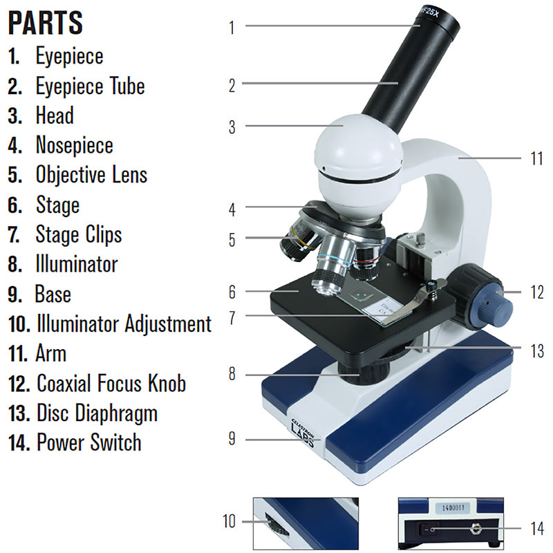
Celestron Labs Compound Microscope CM1000C
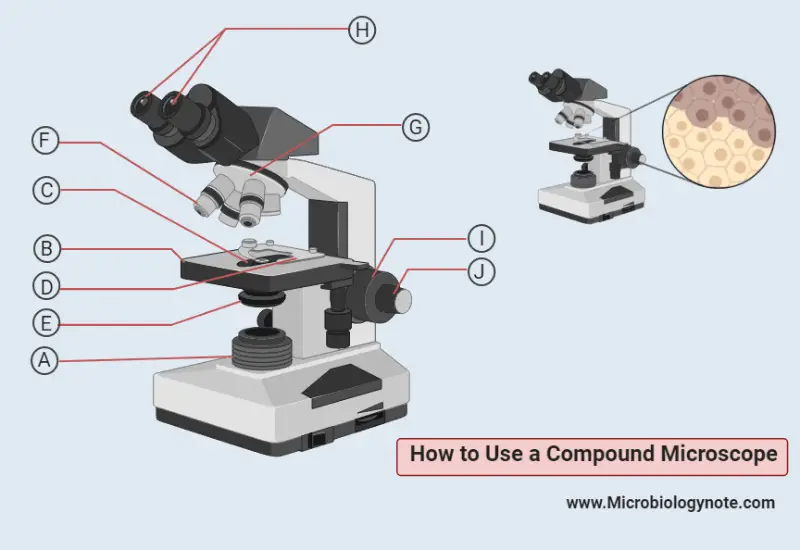
How to Use a Compound Microscope

How To Use a Compound Microscope - YouTube

View Product Photos
Post a Comment for "40 images of compound microscope with labels"