45 diagram of a cell with labels
Animal Cells: Labelled Diagram, Definitions, and Structure Animal Cells Organelles and Functions. A double layer that supports and protects the cell. Allows materials in and out. The control center of the cell. Nucleus contains majority of cell's the DNA. Popularly known as the "Powerhouse". Breaks down food to produce energy in the form of ATP. Label diagram of a cell - Teaching resources 10000+ results for 'label diagram of a cell' Label Parts of a Chemical Equation Diagram Labelled diagram. by Eileenallison
Label Cell Parts | Plant & Animal Cell Activity | StoryboardThat Create a cell diagram with each part of plant and animal cells labeled. Include descriptions of what each organelle does. Click "Start Assignment". Find diagrams of a plant and an animal cell in the Science tab. Using arrows and Textables, label each part of the cell and describe its function.
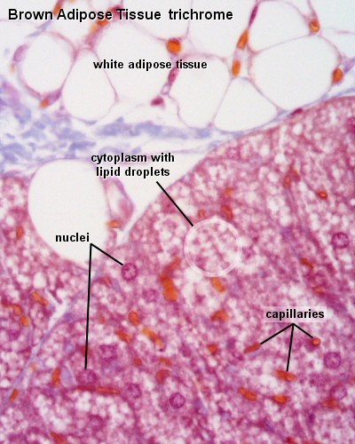
Diagram of a cell with labels
Cell Diagrams - The Biology Corner Open Google Draw and import the diagram. Then use "insert" to create text boxes where students can fill in the labels. Don't forget when assigning this to students on Google classroom to make a copy for each student. You can leave documents in an uneditable form and students can use an addon like "Kami" to annotate the document. How to draw an animal cell - labeled science diagram - YouTube Download a free printable outline of this video and draw along with us: you for watching. Please ... 140,336 Labelled cell Images, Stock Photos & Vectors - Shutterstock Find Labelled cell stock images in HD and millions of other royalty-free stock photos, illustrations and vectors in the Shutterstock collection. Thousands of new, high-quality pictures added every day.
Diagram of a cell with labels. assignmentessays.comAssignment Essays - Best Custom Writing Services Get 24⁄7 customer support help when you place a homework help service order with us. We will guide you on how to place your essay help, proofreading and editing your draft – fixing the grammar, spelling, or formatting of your paper easily and cheaply. Diagram of a cell membrane with labels - NIST Essential Biological FunctionsImmune response, Cell metabolism, Neurotransmission, Photosynthesis, Cell adherence, Cell growth and differentiationPotential Commercial ApplicationsDrug response monitoring, Chemical manufacturing, Biosensing, Energy conversion, Tissue engineering ... Diagram of a cell membrane with labels. Appears In. Biology in ... Plant Cells: Labelled Diagram, Definitions, and Structure Plants have a rigid cell wall that surrounds the plasma membrane. The cell wall is made of cellulose and lignin, which are strong and tough compounds. Plant Cells Labelled Plastids and Chloroplasts Plants make their own food through photosynthesis. Plant cells have plastids, which animal cells don't. Label the cell - Teaching resources - Wordwall by Mbauer. Correctly Label the Bacteria (Prokaryotic) Cell Labelled diagram. by Bronwyn12. Label Plant and Animal Cell Labelled diagram. by Catherine34. Plant Cell - Label Organelles Labelled diagram. by Azimmer. Animal Cell Label Labelled diagram. by Taraabbott.
A Labelled Diagram Of Neuron with Detailed Explanations A Labelled Diagram Of Neuron with Detailed Explanations Biology Biology Article Diagram Of Neuron Diagram Of Neuron A neuron is a specialized cell, primarily involved in transmitting information through electrical and chemical signals. They are found in the brain, spinal cord and the peripheral nerves. A neuron is also known as the nerve cell. Cell Organelles- Definition, Structure, Functions, Diagram In a plant cell, the cell wall is made up of cellulose, hemicellulose, and proteins while in a fungal cell, it is composed of chitin. A cell wall is multilayered with a middle lamina, a primary cell wall, and a secondary cell wall. The middle lamina contains polysaccharides that provide adhesion and allow binding of the cells to one another. Cells Diagram | Science Illustration Solutions - Edrawsoft Cells Diagram Symbols Edraw software offers you lots of symbols used in cells diagram like cell structure, paramecium, squamous cell, cell division, bacteria, cell membrane, eggs, sperm, zygote, an animal cell, SARS, tobacco mosaic, adenovirus, coliphage, herpesvirus, AIDS, pollen, plant cell model, onion tissue, etc. Cells Diagram Examples A Labeled Diagram of the Plant Cell and Functions of its Organelles A Labeled Diagram of the Plant Cell and Functions of its Organelles We are aware that all life stems from a single cell, and that the cell is the most basic unit of all living organisms. The cell being the smallest unit of life, is akin to a tiny room which houses several organs. Here, let's study the plant cell in detail...
03 Label the Cell Diagram | Quizlet Start studying 03 Label the Cell. Learn vocabulary, terms, and more with flashcards, games, and other study tools. PDF Human Cell Diagram, Parts, Pictures, Structure and Functions Diagram of the human cell illustrating the different parts of the cell. Cell Membrane The cell membraneis the outer coating of the cell and contains the cytoplasm, substances within it and the organelle. It is a double-layered membrane composed of proteins and lipids. A Labeled Diagram of the Animal Cell and its Organelles One can observe the golgi apparatus in the labeled animal cell parts diagram. The golgi apparatus is situated near the cell nucleus and besides the stacked sacs, it also contains large number of vesicles. The main function of this golgi complex is to receive the proteins synthesized in the ER and transform it into more complex proteins. Human Cell Diagram, Parts, Pictures, Structure and Functions Diagram of the human cell illustrating the different parts of the cell. Cell Membrane. The cell membrane is the outer coating of the cell and contains the cytoplasm, substances within it and the organelle. It is a double-layered membrane composed of proteins and lipids. The lipid molecules on the outer and inner part (lipid bilayer) allow it to ...
ANAT2241 Connective Tissue Components - Embryology
A Well-labelled Diagram Of Animal Cell With Explanation - Byju's Well-Labelled Diagram of Animal Cell The Cell Organelles are membrane-bound, present within the cells. There are various organelles present within the cell and are classified into three categories based on the presence or absence of membrane. Listed below are the Cell Organelles of an animal cell along with their functions.
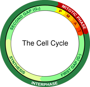
Cell Division
› about › newsroomEsri Newsroom | Publications, Stories, Articles & Press Coverage Jul 08, 2019 · Explore thought-provoking stories and articles about location intelligence and geospatial technology. Discover thought leadership content, user publications & news about Esri.
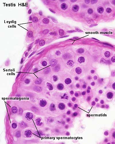
ANAT2341 Lab 1 - Spermatogenesis - Embryology
Animal Cell Diagram with Label and Explanation: Cell Structure, Functions Animal cell is a typical Eukaryotic cell enclosed by a plasma membrane containing nucleus and organelles which lack cell walls, unlike all other Eukaryotic cells. The typical cell ranges in size between 1-100 micrometers. The lack of cell walls enabled the animal cells to develop a greater diversity of cell types.
Talk:ANAT2511 Introduction to Histology - Embryology
en.wikipedia.org › wiki › Flow_cytometryFlow cytometry - Wikipedia Labels, dyes, and stains can be used for multi-parametric analysis (understand more properties about a cell). Immunophenotyping is the analysis of heterogeneous populations of cells using labeled antibodies [35] and other fluorophore containing reagents such as dyes and stains.
Post a Comment for "45 diagram of a cell with labels"